Pet Cherry Eye
A cherry eye is a common term for third eyelid gland prolapses (pop out) due to glandular swelling at medial canthus hyperemia and increased gland volume.
Pet Cherry Eye in Shawnee, OK
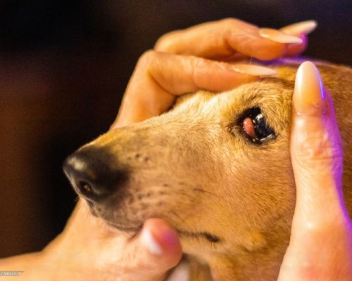
Dogs have an additional eyelid known as the third eyelid (nictitating membrane) that slides across their eyes and is moistened by a gland at its base. Sometimes, it may move out of place and protrude from behind the eyelid, becoming a reddish or pinkish mass at the corner of the eye. It is called a cherry eye because it resembles a cherry sitting at the corner of the eye.
Pet Cherry Eye
Dogs with cherry eyes have a weaker attachment between the third eyelid and the gland. Typically, the gland is firmly attached in place. However, a weaker gland may dislodge the eyelid and bulge out of place. This happens when a tear in the dog’s third eyelid gland leads to inflammation. Medically, a cherry eye is known as a prolapsed lacrimal tear gland of the nictitating membrane or eyelid protrusion. The nictitating membrane acts as a windscreen wiper spreading the tear film evenly across the eyes to keep the cornea healthy and always lubricated. The condition commonly affects young dogs aged between 6 months to 2 years.
Every eye has two glands, one above the eye and the other, the third eyelid, which is lubricated by the tear film made of oil, mucus, and water. The third eyelid gland produces approximately 30% and 60% of tear film water. Therefore, it is crucial to maintain its proper function. Cherry eye causes discomfort, pain, and irritation when inflamed and can affect the function of the third eyelid. The longer the dog stays with the condition, the more irritated the affected eye becomes, which may lead to complications like infections, conjunctivitis, and corneal ulcers. If you have any questions about this condition, please contact our team at Northside Veterinary Hospital!
It is not exactly clear what causes the cherry eye. It is crucial to avoid breeding dogs affected by this condition even if it has already been treated. The condition is primarily seen in the following dog breeds:
- English Bulldogs
- Saint Bernard
- French Bulldogs
- Maltese
- Beagle
- Shih Tzu
- Bulldog
- Lhasa Apso
- Cocker spaniel
- Shar Pei
- Bloodhound
- Pekingese
- Great Dane
- Boston terriers
- Cocker Spaniel
- Shih Tzu
- Pug
- Bullmastiff
- Basset hound
Symptoms of Cherry Eye
Cherry eye may appear suddenly and can quickly progress. The dog may rub the eye leading to bleeding and infection. It is essential to take your dog to the vet for treatment. The most common symptoms include:
- A smooth red or pinkish swollen mass at the corner of the eye near the nose or muzzle
- Irritated, inflamed eye and ulcer
- Swelling around the eye area
- Epiphora (excessive tear production)
- Dog squinting due to pain
- Pus-filled, thick discharge if there is a secondary infection
- Dry eye or cloudy eye due to reduction of tears
- Squinting due to pain
- It may occur in one or both eyes
- Behavior changes like sleeping most of the time to keep the eyelid closed to avoid discomfort and irritation
Causes of Cherry Eye
It is not fully understood why the cherry eye occurs, but when it does, it requires immediate treatment to avoid pain, discomfort, and other eye complications. If the cherry eye is not treated, it may develop dry eye (keratoconjunctivitis). This condition requires daily administration of medicated drops or ointment for life.
One of the causes may be the tear-producing gland becoming displaced from its usual position. Tears moisten the eyes, preventing irritation and supplying nutrition to the cornea to prevent infections. An irritated tear gland struggles to produce tears leading to eye irritation.
Exposure to environmental elements such as dust, microbes, dirt, and the wind may lead to abrasion, secondary infection, and dislocation of the gland. Other factors include:
- Weak ligaments that hold the gland in position may lead to infection in both eyes.
- Genetic predispositions make some dogs, such as brachycephalic breeds (dogs with flat, squished, wide faces and short limbs), have an eye structure that requires stronger tissues and is more prone to the cherry eye than other breeds
- Eye trauma or injury may lead to ligament weakness
- Age; Young puppies have a high likelihood of acquiring the condition
- Abnormal thickening and swelling may prevent the third eyelid from staying in its usual position.
The cherry eye appearance may change in severity if it is improving. However, it is essential to take them to the vet as soon as possible. This will minimize any severe or permanent damage to the third eyelid.
Cherry Eye Diagnosis
The mass growth is easy to identify during diagnosis. Your vet will diagnose your dog by conducting a clinical history and a thorough physical examination. All aspects of the eye will be inspected to check for foreign objects, pus-related infections, and conjunctivitis. Senior dogs will be tested for cancer to rule it out. A fluorescein stain test may also be done in case of scratches in the corneal, which may be infected, perforated, and painful, and if left untreated, it may lead to ulceration.
A Schirmer test is done to test your dog’s tear production to check for dry eyes. It is essential to inform the doctor about any other additional behavior changes in your dog. These test results will enable your vet to devise a treatment plan. A prolapsed third eyelid is a straightforward diagnosis and may not require additional imaging and blood tests.
Cherry Eye Treatment
Immediate treatment is the best way to resolve a cherry eye successfully.
Non-surgical
Dry eye can be treated with medicated ointment or drops several times daily as your vet instructs. Anti-inflammatory drugs also help reduce swelling, and antibiotics and other medications strengthen the ligaments. Downward diagonal toward snout closed eye massages to the affected eye may also help. In some cases, the prolapse may self-correct with manual massage manipulation accompanied by medication.
Surgery
This is the most common method of correcting a cherry eye. Types of surgical procedures include:
Tucking method: It is the most common procedure where the gland is sutured in place and tacked to the orbital rim.
Imbrication method (pocketing): This is a newer technique used when tucking fails. A small portion of tissue in the third eyelid gland is removed then the gap is closed by tiny stitches that tighten the connection between the gland and the third eyelid. Tightening the incision pushes the gland back in position.
After surgery, your dog’s eye will be monitored for normal tear production to ensure the replaced gland has maintained normal functioning.
Cherry Eye Recovery
Surgery has a 90% success rate, but a relapse may occur in rare cases, and a second surgery is performed. The affected eye may appear inflamed for one to two weeks before subsiding. The eye will require ointment application for 7 to 10 days and oral antibiotics for 5 to 10 days.
Your dog must wear an Elizabethan or E collar for a couple of weeks, depending on how fast it recovers and how tempted it is to rub its eye. The collar prevents the dog from damaging or irritating the eye before it recovers and avoids infections. Rechecks are necessary every 2 to 4 weeks unless a complication occurs. Your vet will give you possible signs of difficulty to look out for as the dog recovers. If a complication occurs, a Schirmer Tear test is performed to check whether the dog could have a dry eye which can lead to chronic dry eye.
Cherry eye is not preventable. Therefore, the best way to protect it from complications and relapse is by taking it for regular eye checkups to monitor possible signs of a cherry eye all its life. This is because even with surgery, 10 to 20% of cases of cherry eye relapse even after being surgically corrected.
Please keep your dog healthy by giving it a balanced diet and plenty of exercise to maintain a healthy weight.
For more information about the cherry eye and the procedures that our expert vet surgeons perform to correct it, contact Northside Veterinary Hospital today.

